#lorem ipsum#
Via San Esempio 201, 219038
+ 39 378162183612387612
I am aware that you will use these images for your presentations and I accept it, I would do the same.
At least thank the author of the report and the website
(example: courtesy of AUTHOR(S), see more on www.gastroatlas.org)
Sort by technique (Endoscopy, Ultrasound, etc.) or anathomical site
Free search text (e.g. by author, pathology, anathomical site, etc.)
















1 2 3 4 › 4
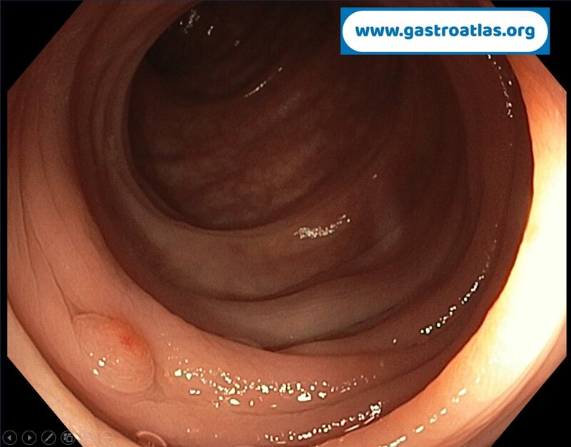
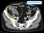
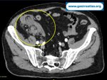
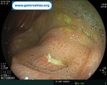
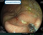
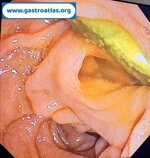
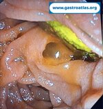
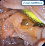
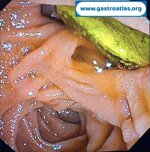
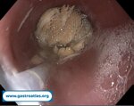
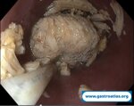
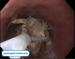
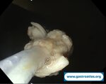
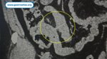
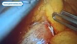
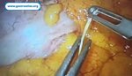
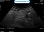
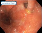
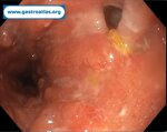
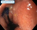
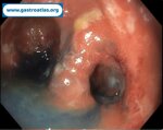
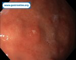
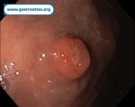
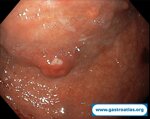
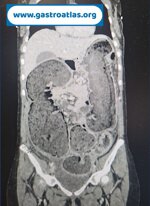
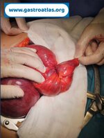
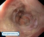
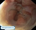
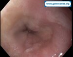
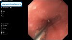
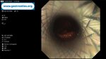
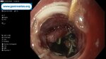
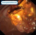
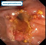
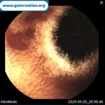
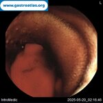
Colonic Inverted Diverticulum (ICD)
2 Endoscopy 1 Clinical case L Colon IBD and other diseases 7 Technique diverticulosis, conti laura, colonic inverted diverticulum (icd), lesion mimicking polyp, colonic perforation
Author: CONTI Laura Roma Italy
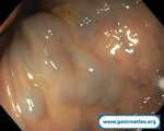
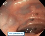
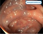
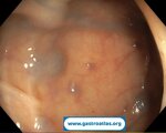
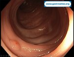
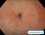
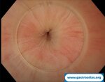
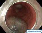
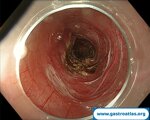
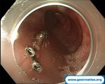
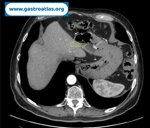
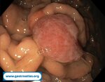
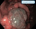
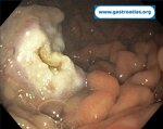
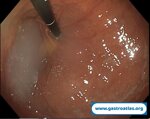
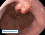
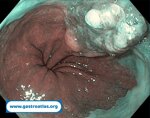
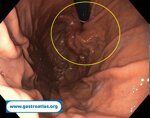
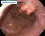
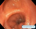
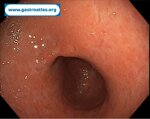
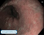
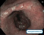
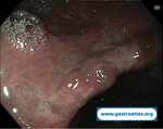
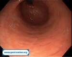
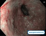
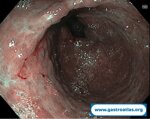
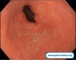
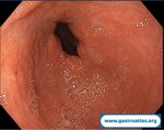
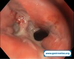
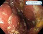
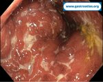
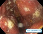
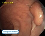
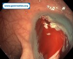
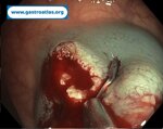
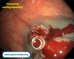
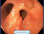
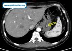
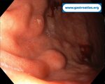
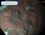
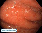
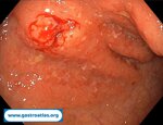
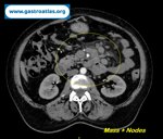
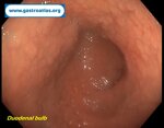
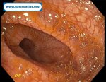
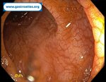
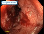
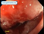
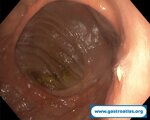
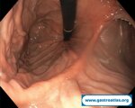
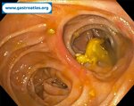
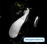
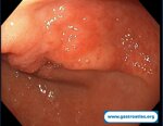
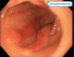
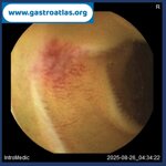
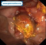
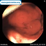
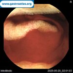
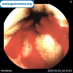
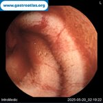
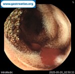
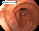
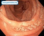
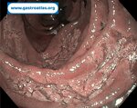
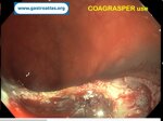
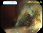
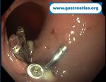
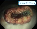
An inverted diverticulum is rare (0.7–1.7% of colonoscopies) but can easily be mistaken for a polyp. Performing a polypectomy or biopsy may cause colonic perforation, a complication well documented in the literature. Recognizing it is essential to avoid hazardous interventions.
During colonoscopy, an inverted diverticulum may show:
Sessile, polyp‑like elevation — similar to a polyp but without a stalk
Fine concentric folds around the lesion (Aurora rings), highly suggestive
Mucosa identical to the surrounding mucosa — no change in pit pattern
Umbilicated or central dimple appearance
Location in diverticular segments — especially the sigmoid colon (≈75%)
Dynamic signs that help distinguish an ICD from a true polyp:
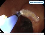
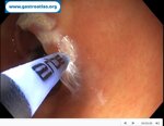
Eversion with biopsy forceps — gentle pressure may cause the lesion to “pop back” into the diverticular lumen
Water or air insufflation — the lesion may collapse and reveal a typical diverticulum
Methylene blue staining — highlights the concentric folds